The lateral cervical flap comprising skin fascia and platysma muscle has significant applications in the head and neck region after radical ablative surgery for cancer of the oral cavity.
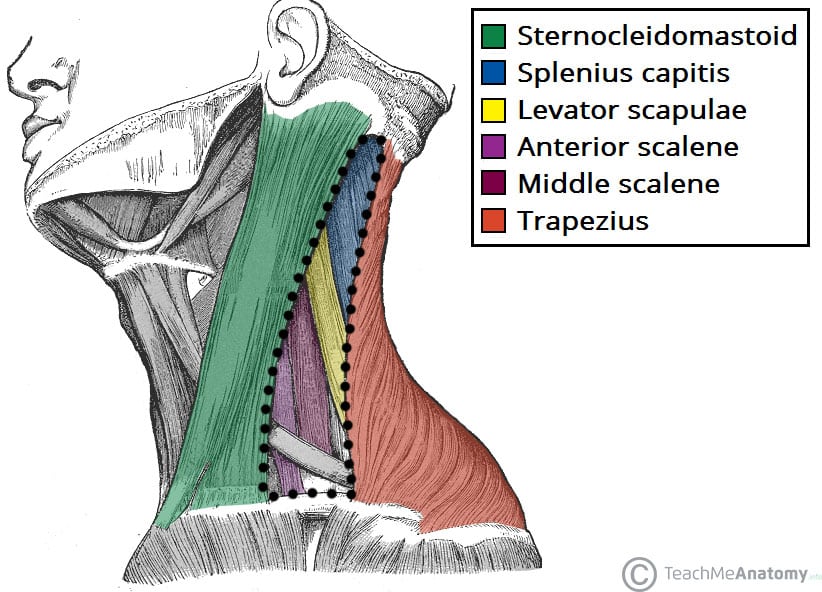
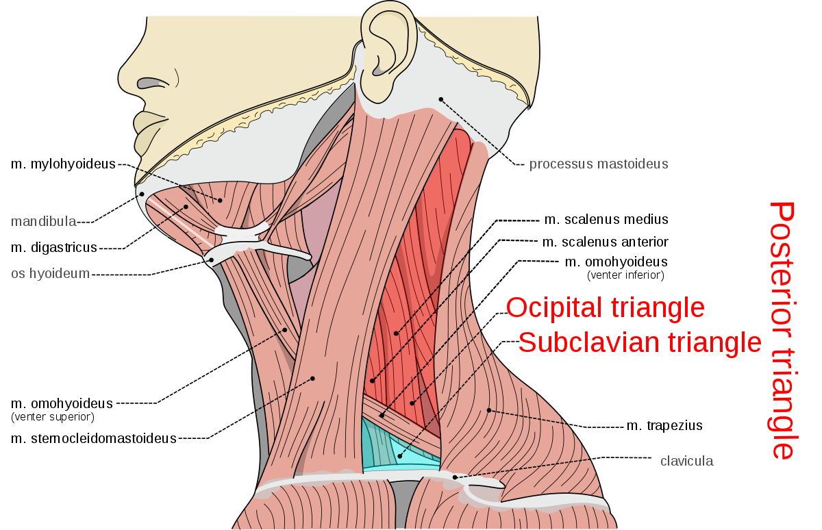
Floor of lateral cervical region.
The posterior triangle of the neck is covered by the investing layer of fascia and the floor is formed by the prevertebral fascia see fascial layers of the neck.
Posterior cervical region lateral cervical region anterior cervical region.
The anterior triangle is a region located at the front of the neck.
Lateral cervical region the lateral cervical region has three main borders.
In this article we shall look at the anatomy of the anterior triangle of the neck its borders contents and subdivisions.
They can be divided into anterior lateral and posterior groups based on their position in the neck.
The muscles in the neck are responsible for the movement of the head in the cervical region in all directions.
This region includes lots of muscles nerves arteries and veins and lymph.
The posterior triangle or lateral cervical region is a region of the neck boundaries.
Union of the.
It courses posteroinferiorly along the floor of the posterior triangle from the scm and disappears by coursing deep to the which it also.
Its boundaries are as follows.
As shown in the figure above the region is inferior to the mandible anterior to the internal jugular vein and superior to the clavicle.
Anterior posterior border of the sternocleidomastoid.
Investing layer of the deep cervical fascia.
They are further divided into more specific groups based on a number of determinants.
They are located on both the left and the right sides of the neck.
Posterior anterior border of the trapezius muscle.
Inferior middle 1 3 of the clavicle.
The mandible the internal jugular vein and the clavicle.
From superior to inferior 1 m.
The musculature of the neck is comprised of a number of different muscle groups.
The is a key landmark of the neck as it divides the anterior cervical region from the lateral cervical region.


