Inferiorly hyoid bone.
Floor of carotid triangle is formed by.
It is so called because it contains all the 3 carotid arteries viz.
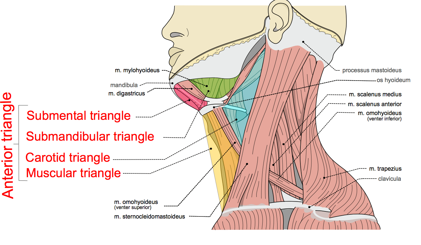
The triangles of the neck are the topographic areas of the neck bounded by the neck muscles.
What muscles form the floor of the posterior triangle.
Using the digastric and omohyoid muscles it is common to divide the anterior triangle into smaller submandibular submental carotid and muscular triangles to descriptive purposes.
Shahab shahid mbbs reviewer.
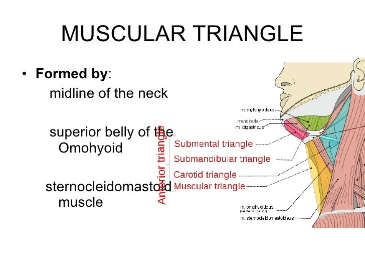
Superior belly of omohyoid.
Common carotid internal carotid and external carotid its boundaries are.
Posterior belly of digastric and stylohyoid.
Its floor is formed by parts of the thyrohyoid membrane hyoglossus and the.
Constrictores pharyngis medius and inferior.
Medially midline of the neck.
The triangles of the neck are important because of their contents as they house all the neck structures.
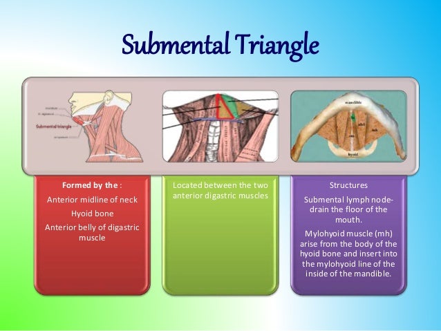
It contains the submental lymph nodes which filter lymph draining from the floor of the mouth and parts of the tongue.
Uruj zehra mbbs mphil phd last reviewed.
August 31 2020 the neck or cervical region is perhaps one of the most anatomically complex regions of the body despite being a relatively small region the contents within this region and notably the interrelationships between them hold a great deal of anatomical functional and.
Floor formed by the pharynx.
Name the structures forming the boundaries of carotid triangle.
The sternocleidomastoid muscle divides the neck into the two major neck triangles.
The external and internal carotids lie side by side the external being the more anterior of the two.
The carotid triangle also contains.
Common carotid artery internal jugular vein vagus nerve and hypoglossal nerve.
From anterior to posterior scalenus anterior scalenus medius levator scapulae splenius capitis.
Floor of the anterior cervical triangle the floor of the anterior triangle of the neck is formed mainly by the pharynx larynx and thyroid gland.
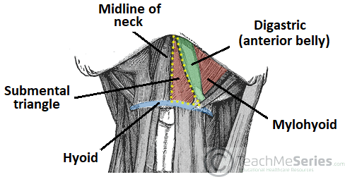
Laterally anterior belly of the digastric.
The common carotid artery bifurcates within the carotid triangle to form the external and internal carotid arteries.
Hypoglossal nerve is a content of both digastric carotid triangles.
The anterior triangle and the posterior triangle of the neck each of them containing a few subdivisions.
List the important structural contents of the carotid triangle.
Carotid triangle is one of the subdivisions of anterior triangle of neck.
The base of the submental triangle is formed by the.
Structure superficial to mylohyoid in anterior digastric triangle is mylohyoid artery nerve.
Floor of digastric triangle is formed by mylohyoid anteriorly hyoglossus posteriorly infrahyoid ribbon muscles are the chief contents of muscular triangle.
This muscular triangle actually has four sides and is situated more inferiorly than the other triangles.